
The exocciput and supraocciput were smaller and the tentorium was steeper, but there was no difference in posterior fossa brain volume. (1997) found that 30 patients with Chiari type I malformation had a significantly smaller bony posterior fossa compared to controls. (1941) have been credited with the first detailed clinical description of Chiari type I malformation, although confusion regarding the nomenclature existed up to the 1970s ( Schijman, 2004). Caudal migration of the brainstem and cerebellar tonsils often associated with syringomyelia but without spina bifida has been referred to as 'Chiari type 1.5' ( Schijman, 2004 Tubbs et al., 2004).Īdams et al. These patients have resolution of symptoms with decompression surgery ( Iskandar et al., 1998 Tubbs et al., 2001). 'Chiari type 0' has been defined as syringohydromyelia without cerebellar tonsillar herniation, but with distortion of the contents of the posterior fossa.

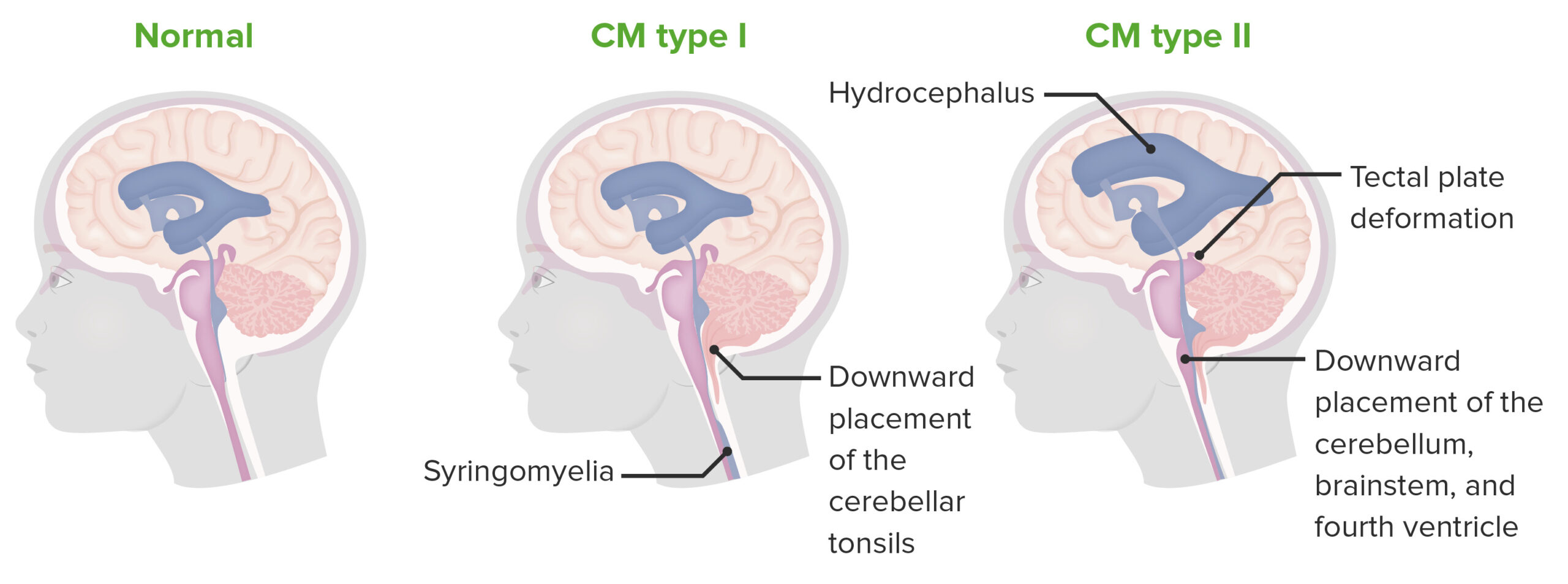
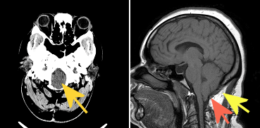
Two additional types of Chiari malformation have been described. Whereas Chiari types II, III, and IV are considered to be primarily neural in origin resulting from neuroectodermal anomalies, Chiari type I is considered to result from a mesodermal defect ( Schijman, 2004). Type IV refers to hypoplasia or aplasia of the cerebellar hemispheres and alterations of the pons with marked dilatation of the fourth ventricle, cisterna magna, and basal cisterns. Hydrocephalus is associated in 50% of cases and is due to aqueductal stenosis or Dandy-Walker malformation. Part of the hindbrain may also be herniated. Type III is characterized by caudal displacement of the medulla and herniation of part of the cerebellum in an occipital or cervical meningocele. Type II is the most frequent form of Chiari malformation and is associated with spina bifida (see 182940) and hydrocephalus. In Chiari type II (CM2 207950), there is descent of the cerebellar tonsils, cerebellar inferior vermis, and portions of the cerebellar hemispheres into the spinal canal along with displacement of the brainstem and fourth ventricle. Type I is descent of the cerebellar tonsils into the cervical canal and is usually not associated with hydrocephalus. Syringomyelia, noncommunicating (80% of cases) ĭecreased volume of the posterior cranial fossa with normal hindbrain volume ĭecreased CSF volume in posterior fossa įour classic types of Chiari malformation have been described. Herniation of the cerebellar tonsils through the foramen magnum 5 mm or greater Headache, suboccipital, migraine-like (most common symptom) precipitated by coughing, sneezing, bending forward, lifting, neck extension Įxtensor plantar responses Ĭhiari I malformation on MRI Segmental sensory loss, especially of pain and temperature

Touch, vibration, and limb position may or may not be affected Loss of pain and temperature in a cape-like distribution

Neck pain Īrm pain īurning pain in the limbs Hyperreflexia, especially of the lower limbs Īreflexia of the upper limbs


 0 kommentar(er)
